Small animal X-ray – what went wrong?
Look below at this article: Small animal X-ray Mistakes?
A clinical article explaining the best techniques that can be used to obtain the best quality images from your radiographs.
Small animal X-ray – What went wrong?
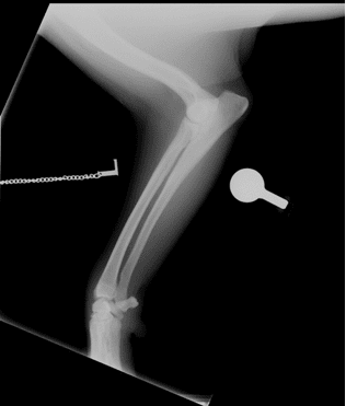
At first glance this image is a little pale but not too bad. There is a marker, and the view is fairly lateral. There is also an orthopaedic calibration ball.
However, as you look closer it is possible to see that the image is in fact very poor quality.
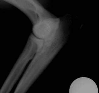
There is no bony trabecular detail visible, resulting in an image that is non-diagnostic, particularly around the elbow joint.
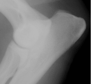
Even after using the abilities of the CR system to darken the image there is still no detail visible within the bone.
To improve this image, we definitely need more mAs. Darkening and magnifying the image reveals quantum mottle over the thicker regions, which clearly indicates that the mAs is too low.
Put simply, you need X-ray photons to make a good radiograph!
But also it is very hard to correctly position and expose an image to show both the carpus and elbow. The best thing to do is perform 2 separate radiographs ensuring both are correctly exposed.
The main reason for attempting a ‘forearm’ radiograph is to look for bony alignment.
So to improve this image my advice would be:
- Either radiograph the elbow or the carpus.
- Increase the mAs.
- Collimate a bit tighter to reduce scatter.
One common mistake when radiographing extremities is to use too little mAs and to set the kV too high. This does have the benefit of reducing the risk of motion. But the fundamental issue then is that you will lack bony trabecular detail, and will end up with reduced image quality. You really do need radiation to form a decent image. No matter how good the post-processing on a CR or DR image you do still need X-Ray photons!
I often say to think of using a digital camera in a dark room. You get a noisy grainy image due to a lack of photons. A noisy, grainy radiograph is a lack of X-ray particles.
The correct name for an image that is grainy and lacking data is quantum mottle.
One definition is that quantum mottle is a type of radiographic noise directly related to the number of X-ray photons exiting the patient. Forming the radiographic image; fewer photons reaching the detector will cause an undesirable fluctuation in image densities, resulting in images with a grainy or sand-like appearance.